Newport Eye Center - Office Views
Newport Eye Center - Office Views
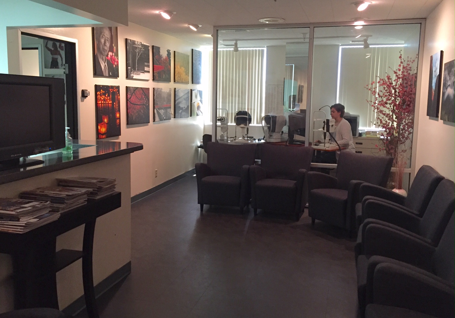
Waiting room with screening area beyond.
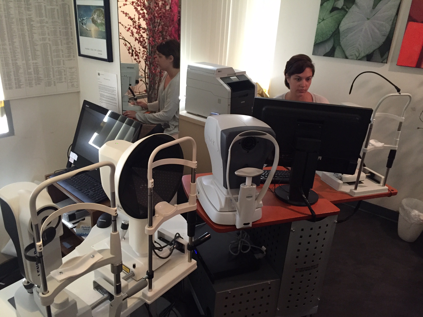
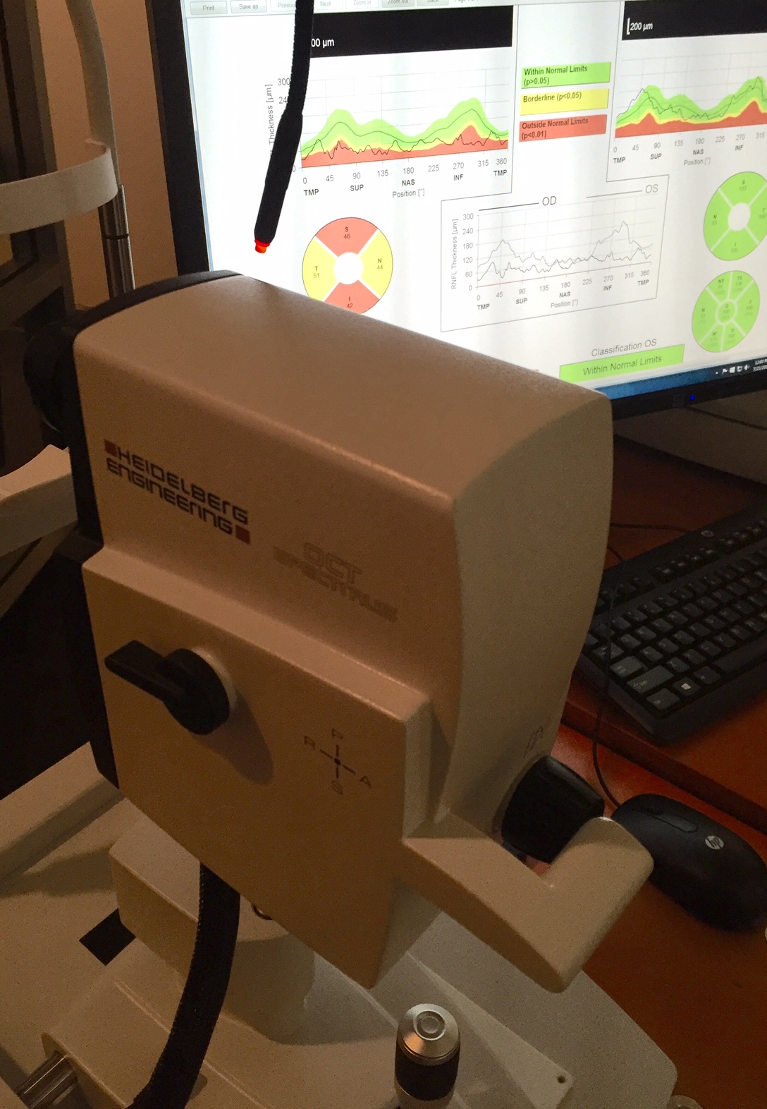
This area contains the Heidelberg Spectralis OCT, which scans at 84,000 images/sec, the Haag-Streit Lenstar and Cassini topographer.
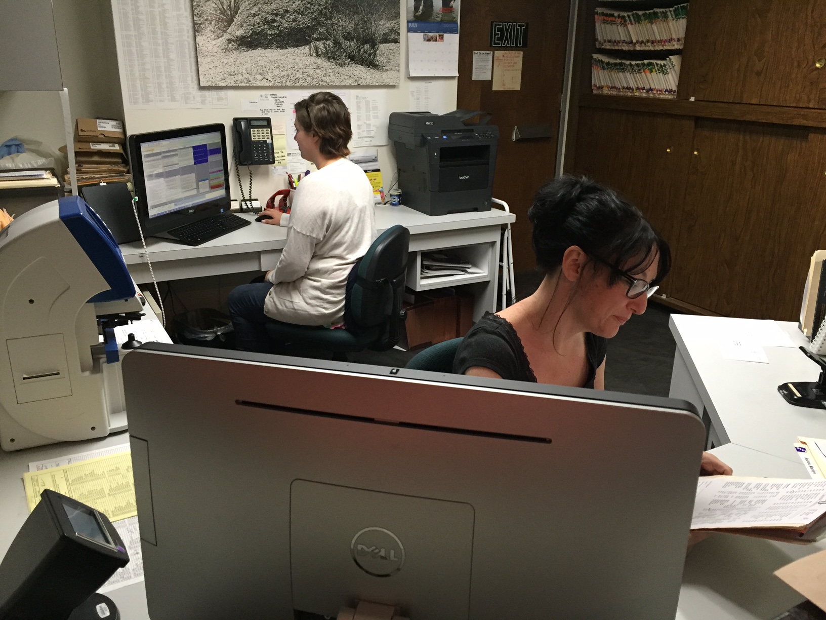
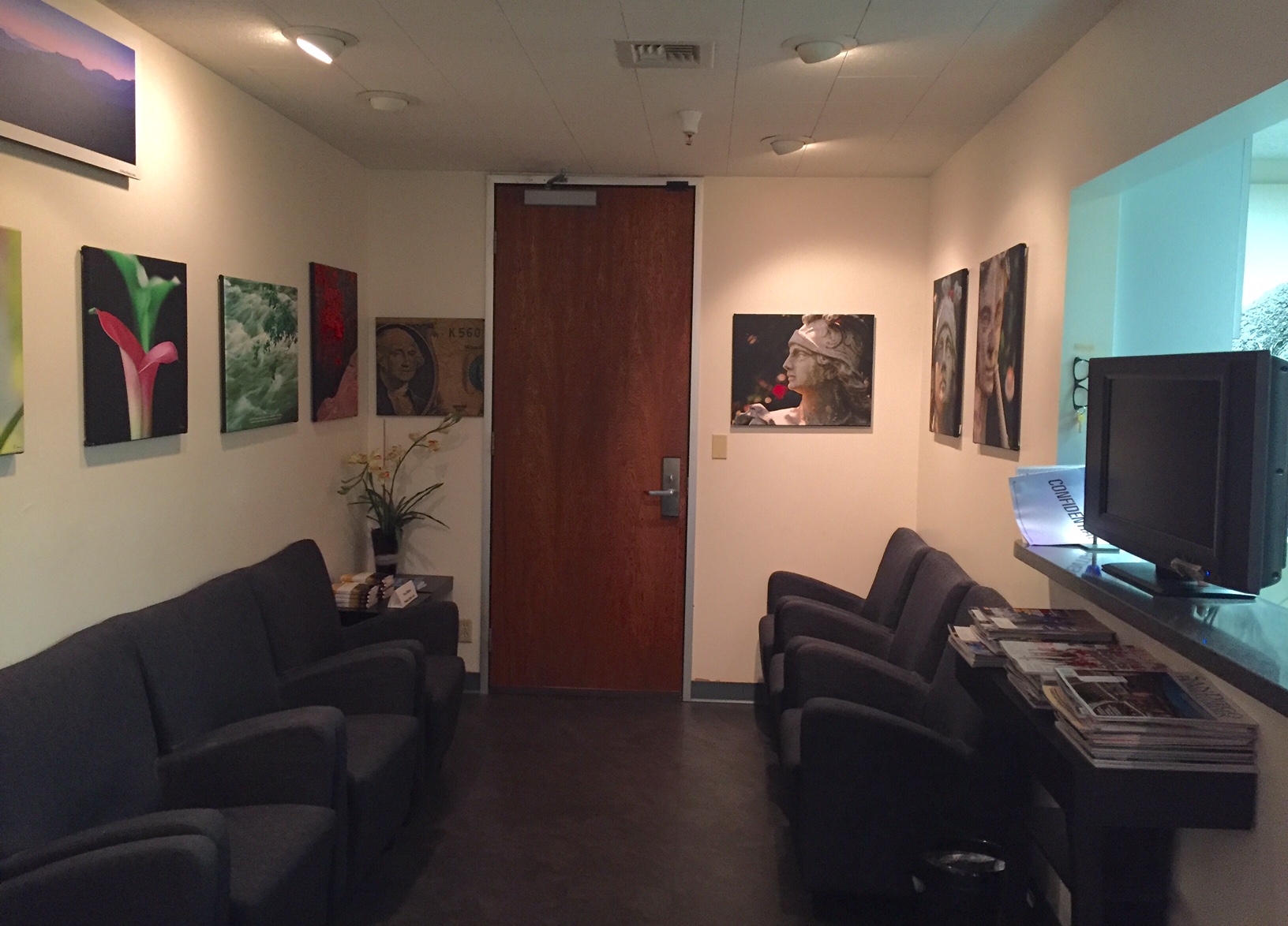
Entrance and reception areas.
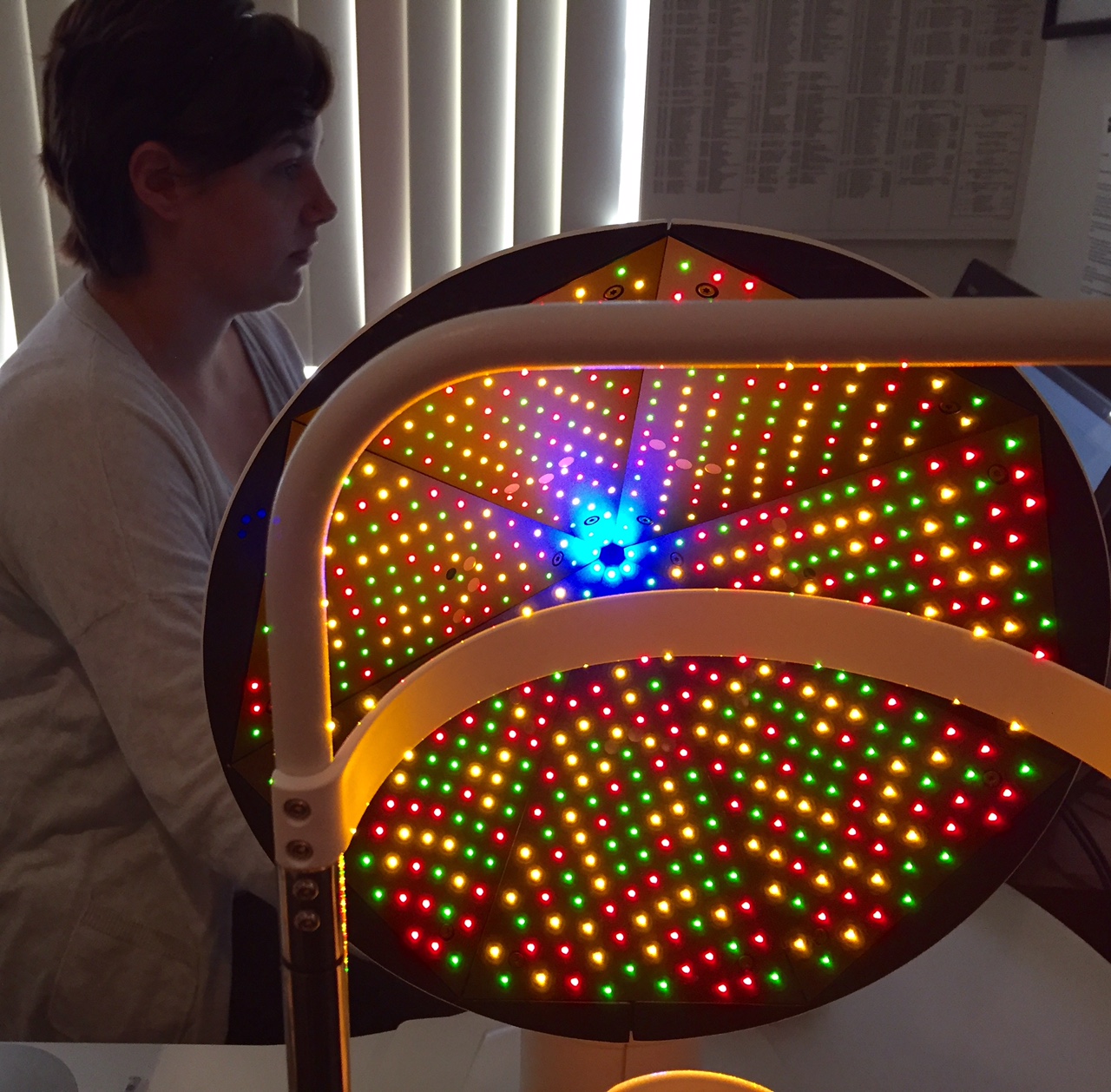
The "Christmas Tree" lights that provide anterior and posterior corneal curvatures for more accurate IOL determinations using customized software developed in-house.
Glaucoma results in background on display: severe loss of nerve fibers on left side (red) and normal findings on right side (green).